
Comprehensive Guide to Nail Diseases: Understanding Common Conditions and Systemic Manifestations
The nail unit serves as more than just a protective structure for our fingertips and toes—it functions as a diagnostic window into our overall health. According to dermatological research and clinical observations, nail abnormalities can indicate local pathology, systemic diseases, nutritional deficiencies, and even psychological conditions. This comprehensive guide explores the most common nail diseases, their clinical manifestations, diagnostic approaches, and treatment strategies based on current medical understanding.
Understanding the Nail Unit Architecture:
The nail unit comprises four essential components that work together to produce and maintain healthy nails. The nail plate, composed primarily of keratin protein, water, lipids, and trace elements, represents the visible portion we commonly refer to as the nail. This structure grows at approximately 3mm per month for fingernails and 1mm per month for toenails, serving aesthetic, protective, and functional purposes including object manipulation and fine motor tasks.
The nail matrix, located beneath the proximal nail fold, acts as the growth center where new nail cells are produced. The nail bed provides support and nutrition to the nail plate, while the nail folds (proximal and lateral) create protective barriers around the nail edges. Understanding this anatomy is crucial for recognizing how various diseases affect different components of the nail unit.
Fungal Infections: The Most Common Nail Pathology
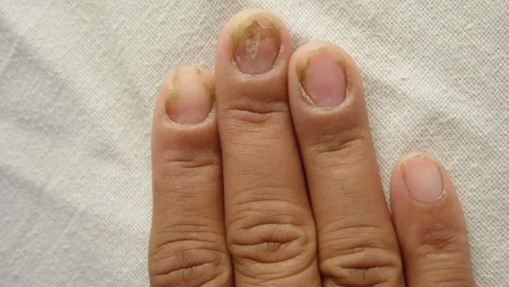
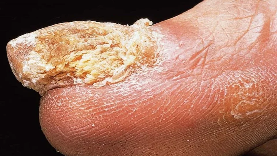
Onychomycosis, or fungal nail infection, represents the most prevalent nail disorder, affecting approximately 10-20% of the global population according to epidemiological studies. The World Health Organization recognizes this condition as a significant public health concern due to its chronic nature and impact on quality of life.
Etiology and Risk Factors
Two primary fungal groups cause onychomycosis: dermatophytes (most common) and Candida species. Dermatophytes, particularly Trichophyton rubrum and Trichophyton mentagrophytes, account for approximately 80-90% of toenail infections and 50% of fingernail infections. Risk factors include advancing age, diabetes mellitus, immunocompromised states, peripheral vascular disease, and repetitive nail trauma.
Clinical Manifestations:
Fungal infections typically present with characteristic features including yellow-brown discoloration of the nail plate, subungual hyperkeratosis (thickening of tissue beneath the nail), onycholysis (separation of nail plate from nail bed), and in severe cases, complete nail destruction. The infection often begins at the distal or lateral nail edge and progressively spreads toward the nail matrix.
Diagnostic Approaches:
Accurate diagnosis requires laboratory confirmation through potassium hydroxide (KOH) preparation and fungal culture. Direct microscopic examination of nail clippings treated with KOH can immediately identify fungal elements, while culture remains the gold standard for species identification and antifungal sensitivity testing.
Treatment Strategies:
Current treatment guidelines recommend systemic antifungal therapy for most cases. Itraconazole pulse therapy (400mg daily for 7 days per month, with 2 pulses for fingernails and 3 pulses for toenails) and Terbinafine continuous therapy (250mg daily for 6 weeks for fingernails and 12 weeks for toenails) represent first-line treatments with cure rates ranging from 60-80% according to clinical trials.
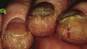
Paronychia: Inflammation of the Nail Folds:
Paronychia, characterized by inflammation of the nail folds, presents in both acute and chronic forms, each requiring different therapeutic approaches.
Acute Paronychia
Acute paronychia typically results from Staphylococcus aureus infection following minor trauma such as aggressive manicuring, hangnails, or ingrown nails. Clinical presentation includes painful, red swelling of the nail fold, often with purulent discharge. Treatment involves systemic antibiotics (typically anti-staphylococcal agents) and surgical drainage when pus collection is present.
Chronic Paronychia
Chronic paronychia predominantly affects individuals with chronically wet hands, such as healthcare workers, food handlers, and cleaning personnel. Candida albicans frequently colonizes the chronically inflamed tissue, leading to persistent swelling, cuticle loss, and transverse nail grooves. Management focuses on moisture protection, topical antifungals, and in severe cases, oral antifungal therapy with fluconazole or itraconazole.
Eczematous Nail Changes:
Nail involvement in eczema affects 10-15% of patients with chronic hand dermatitis according to dermatological studies. Both irritant and allergic contact dermatitis can significantly impact nail morphology.
Common Triggers:
Frequent exposure to rubber, chromium compounds, detergents, soaps, and various chemicals can trigger eczematous nail changes. Atopic dermatitis patients show increased susceptibility to nail involvement during disease flares.
Clinical Features:
Eczematous nail changes include nail plate thickening, transverse ridges and grooves (Beau’s lines), subungual hyperkeratosis, and onycholysis. The “sandpapered nail” appearance (trachyonychia) represents a characteristic finding in chronic cases.
Management:
Treatment emphasizes allergen avoidance, protective measures including cotton gloves under rubber gloves, regular application of emollients, and potent topical corticosteroids during acute flares.
Psoriatic Nail Disease:
Nail psoriasis affects 25-50% of patients with cutaneous psoriasis and up to 80% of those with psoriatic arthritis, according to rheumatological research. The condition can precede skin manifestations in some patients, making nail examination crucial for early diagnosis.
Characteristic Features:
Nail pitting represents the most common manifestation, appearing as small, punctate depressions on the nail surface. Other features include oil drop discoloration, subungual hyperkeratosis, onycholysis, and transverse ridging. The severity often correlates with joint involvement in psoriatic arthritis.
Treatment Approaches:
Management ranges from topical therapies (corticosteroids, calcipotriol) for mild cases to systemic treatments including methotrexate and cyclosporin for severe disease. Newer biological therapies show promising results in clinical trials for refractory cases.
Trauma-Related Nail Disorders:
Various forms of trauma can significantly impact nail health and appearance, often requiring specific management strategies.
Nail Biting and Habit-Tic Deformity:
Chronic nail biting affects approximately 20-30% of children and 5% of adults according to behavioral studies. Habit-tic deformity, caused by repetitive manipulation of the proximal nail fold, creates characteristic transverse ridges with a central longitudinal groove resembling a “fir tree” pattern.
Footwear-Related Trauma:
Ill-fitting shoes can cause onychogryphosis (ram’s horn nail deformity) and ingrown toenails. These conditions are particularly common in elderly populations and athletes, requiring both conservative management and sometimes surgical intervention.
Nail Manifestations of Systemic Diseases
The nail unit serves as an important diagnostic tool for recognizing systemic conditions, with specific patterns correlating with different disease states.
Cardiovascular and Pulmonary Conditions:
Clubbing, characterized by increased nail curvature and soft tissue hypertrophy, indicates chronic hypoxemia associated with cardiovascular, bronchopulmonary, and gastrointestinal disorders. Early recognition can prompt investigation for underlying pathology.
Nutritional and Metabolic Disorders:
Koilonychia (spoon-shaped nails) strongly suggests iron deficiency anemia, while Muehrcke’s lines (white bands) indicate hypoalbuminemia. According to hematological studies, these findings often precede laboratory abnormalities.
Renal Disease:
Half-and-half nails, showing proximal pallor with distal reddish-brown discoloration, characteristically appear in chronic renal failure patients and may correlate with disease severity.
Endocrine Disorders:
Thyrotoxicosis frequently causes onycholysis, particularly affecting the ring finger nail. Pregnancy-related hormonal changes can also trigger nail separation in susceptible individuals.
Drug-Induced Nail Changes:
Various medications can cause characteristic nail abnormalities. Cytotoxic drugs commonly produce transverse pigmented bands and growth arrest lines, while antimalarials like chloroquine can cause bluish-black pigmentation. Tetracyclines may trigger onycholysis, particularly with sun exposure.
Diagnostic Considerations and Clinical Examination
Proper nail examination requires systematic evaluation of all components including the nail plate, folds, and surrounding skin. Dermoscopy has emerged as a valuable tool for detecting subtle changes not visible to the naked eye. When systemic disease is suspected, appropriate laboratory investigations including complete blood count, metabolic panels, and specific disease markers should be considered.
Prevention and General Care
Maintaining optimal nail health requires attention to basic hygiene principles including regular but gentle cleaning, appropriate moisturization, avoiding trauma, and using protective equipment when handling chemicals or performing manual labor. Patient education about proper nail care significantly reduces the risk of various nail disorders.
Conclusion
Understanding nail diseases requires comprehensive knowledge of normal nail anatomy, recognition of pathological changes, and awareness of systemic associations. As medical research continues to evolve, new diagnostic techniques and treatment modalities are improving outcomes for patients with nail disorders. The nail unit truly serves as a “window” into both local and systemic health, making its examination an essential component of comprehensive medical evaluation.
Early recognition and appropriate treatment of nail diseases from qualified dermatologist not only improve cosmetic outcomes but can also prevent progression to more serious complications and help identify underlying systemic conditions that might otherwise go undiagnosed.