
Decoding Your Skin: A Comprehensive Guide to Common Dermatological Conditions
Your skin serves as your body’s first line of defense and largest organ, yet many people struggle to understand what various skin changes mean. This comprehensive guide will help you recognize common dermatological conditions, understand their underlying causes, and make informed decisions about treatment options.
Understanding the Language of Skin Lesions
Before exploring specific conditions, it’s crucial to understand how dermatologists classify and describe skin abnormalities. This standardized terminology forms the foundation of accurate diagnosis and effective treatment.
Primary Skin Lesions: The Building Blocks of Diagnosis
Flat Lesions:
- Macule: A flat, discolored area less than 1 centimeter in diameter. Examples include freckles and flat moles. These represent localized color changes without any elevation above the surrounding skin.
- Patch: Similar to a macule but larger than 1 centimeter. Café-au-lait spots and large birthmarks fall into this category.
Raised Lesions:
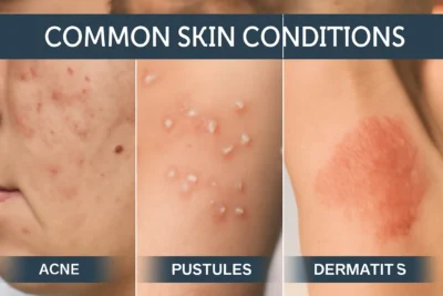
- Papule: A small, solid elevation less than 1 centimeter in diameter. Acne lesions, insect bites, and small warts are common examples.
- Plaque: A raised, flat-topped lesion larger than 1 centimeter. Psoriasis typically presents with characteristic plaques.
Fluid-Filled Lesions:
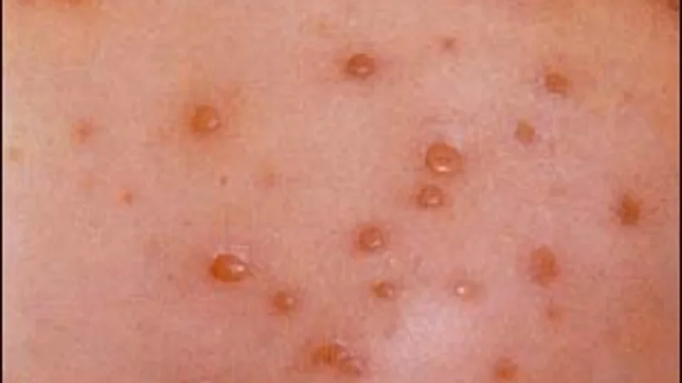
- Vesicle: A small, fluid-filled bump less than 1 centimeter. Chicken pox and herpes simplex create typical vesicular eruptions.
- Bullae: Large fluid-filled lesions greater than 1 centimeter, often seen in severe burns or autoimmune blistering diseases.
Deep Lesions:
- Nodule: A solid, palpable lesion that extends into deeper skin layers, having measurable length, width, and depth.
Understanding these basic morphologies helps in recognizing patterns and communicating effectively with healthcare providers. When documenting skin changes, note the size, color, texture, and distribution of lesions.
Bacterial Skin Infections: Recognition and Management
Bacterial infections represent one of the most common categories of skin disease, ranging from superficial annoyances to serious medical emergencies requiring immediate intervention.
Impetigo: The Highly Contagious Surface Infection
Impetigo stands out as the most frequently encountered bacterial skin infection, particularly affecting children. This superficial infection involves only the outermost layers of skin but spreads rapidly through direct contact.
Clinical Presentation: The condition begins as small vesicles on an erythematous base, which rupture easily due to their thin walls. The resulting serum dries to form the characteristic honey-colored or golden-yellow crusts that define this condition. These lesions heal without scarring, and constitutional symptoms are rare unless secondary complications develop.
Causative Organisms: Two primary bacterial species cause impetigo: Streptococcus pyogenes and Staphylococcus aureus. The relative prevalence of each organism varies geographically and temporally, with important implications for antibiotic selection.
Treatment Strategies:
Topical Therapy (for localized disease):
- Mupirocin 2% ointment: Apply three times daily for 5-7 days. This antibiotic specifically targets gram-positive bacteria and achieves excellent skin penetration.
- Fusidic acid 2% cream: An alternative with similar efficacy, particularly useful in areas with mupirocin resistance.
Systemic Therapy (for extensive disease or treatment failures):
- Erythromycin
- Flucloxacillin
- Clindamycin
Prevention and Control: Proper hygiene, immediate treatment of new cases, and isolation of affected individuals until 24 hours after starting effective antibiotic therapy help control spread in communities and families.
Folliculitis: When Hair Follicles Become Inflamed
Folliculitis occurs when hair follicles become inflamed, typically due to bacterial infection, though mechanical, chemical, or fungal causes also exist.
Clinical Recognition: The condition presents as dome-shaped papules and pustules centered around hair follicles. The distribution often provides clues to the underlying cause – beard area involvement suggests shaving-related trauma, while widespread distribution might indicate systemic factors.
Common Precipitating Factors:
- Chemical irritants from cosmetics or occupational exposures
- Mechanical trauma from tight clothing or adhesive materials
- Occlusion from topical products, including inappropriate steroid use
- Hot, humid environments that promote bacterial overgrowth
Treatment Approach:
Mild Cases:
- Antiseptic washes: Chlorhexidine or benzoyl peroxide cleansers used twice daily
- Topical antibiotics: Clindamycin 1% solution or erythromycin 2% gel applied twice daily
Moderate to Severe Cases:
- Oral antibiotics: Doxycycline or minocycline
- Alternative: Cephalexin for patients who cannot take tetracyclines
Management Success Factors: Identifying and eliminating predisposing factors is crucial for preventing recurrence. This might involve changing shaving techniques, avoiding tight clothing, or modifying occupational practices.
Furuncles: Deep-Seated Hair Follicle Infections
Furuncles represent a more serious progression of follicular infection, extending deep into surrounding tissues and often requiring more aggressive management.
Pathophysiology and Presentation: These infections begin as inflammatory follicular nodules that become increasingly painful and develop central necrosis. The natural history involves progression through inflammatory, pustular, and necrotic phases before eventual healing, often with scarring.
Comprehensive Treatment Protocol:
Conservative Management:
- Warm compresses: Apply for 15-20 minutes, 3-4 times daily to promote drainage and provide symptomatic relief
- Pain management: NSAIDs like ibuprofen address both pain and inflammation
- Activity modification: Avoid manipulation or squeezing, which can spread infection
Antibiotic Therapy:
- First-line: Flucloxacillin oral
- Alternatives: Clindamycin oral
- Severe cases: May require intravenous antibiotics or surgical drainage
Monitoring and Follow-up: Patients should be monitored for signs of spreading infection, including lymphangitis, cellulitis, or systemic symptoms. Failure to respond to appropriate antibiotic therapy within 48-72 hours warrants reevaluation.
Ecthyma: The Deeper Variant
Ecthyma represents a deeper form of pyogenic infection, typically occurring in patients with compromised hygiene or nutritional status.
Clinical Features: Unlike impetigo, ecthyma penetrates through the epidermis into the dermis, creating indurated lesions with thick, adherent crusts. The base shows induration with surrounding erythema, and healing invariably results in scarring.
Treatment Strategy:
- Address underlying factors: Improve hygiene and nutritional status
- Systemic antibiotics: Usually required due to the depth of infection
- Wound care: Gentle debridement and topical antiseptics
Cellulitis: The Spreading Emergency
Cellulitis involves inflammation of subcutaneous tissues and represents a potentially serious condition requiring prompt recognition and treatment.
Clinical Recognition: The hallmark features include diffuse erythema, warmth, swelling, and tenderness. The affected area typically has poorly defined borders that may advance rapidly. Severe cases can develop bullae, necrosis, or systemic toxicity.
Risk Factors:
- Preceding trauma or skin breakdown
- Venous insufficiency or lymphedema
- Diabetes mellitus or immunocompromise
- Previous episodes of cellulitis
Treatment Protocol:
Outpatient Management (mild cases):
- Penicillin V:
- Alternative: Erythromycin for penicillin-allergic patients
- Supportive care: Limb elevation, rest, analgesics
Inpatient Management (severe cases):
- Intravenous antibiotics: Benzylpenicillin plus flucloxacillin
- Monitoring: For systemic toxicity and response to therapy
- Complications: Watch for necrotizing fasciitis or sepsis
Viral Skin Infections: Understanding Persistent Pathogens
Viral infections present unique challenges due to their tendency to establish latency, cause recurrent episodes, and show limited response to conventional antimicrobial therapy.
Herpes Simplex: The Recurrent Challenge
Herpes simplex labialis affects a significant portion of the population and demonstrates the typical pattern of viral skin infections: initial infection followed by periods of latency and episodic reactivation.
Clinical Course: The infection progresses through predictable stages: prodromal tingling, vesicle formation, ulceration, crusting, and healing. The entire cycle typically spans 10-14 days, with constitutional symptoms more common during primary infections.
Treatment Options:
Topical Therapy:
- Acyclovir 5% cream
- Penciclovir 1% cream
- Docosanol 10% cream
Systemic Therapy:
- Valacyclovir, or acyclovir 400mg
Varicella: The Universal Childhood Experience
Varicella, commonly known as chickenpox, typically represents a benign childhood illness but can cause serious complications in adults and immunocompromised individuals.
Clinical Progression: The disease begins with constitutional symptoms followed by the characteristic vesicular eruption appearing in crops. New lesions continue to appear for 3-5 days, with different stages of lesions present simultaneously – a key diagnostic feature.
Treatment Approach:
Healthy Children:
- Symptomatic care: Antihistamines for pruritus, acetaminophen for fever
- Comfort measures: Cool baths, loose clothing, calamine lotion
- Complication prevention: Avoid aspirin due to Reye’s syndrome risk
High-Risk Patients:
- Acyclovir
- Monitoring: For complications including pneumonia, encephalitis, or secondary bacterial infections
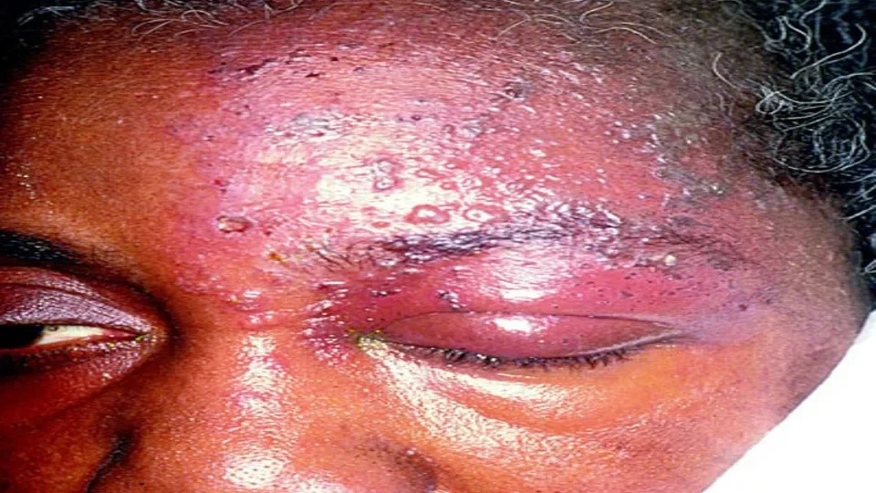
Herpes Zoster: The Reactivation Syndrome
Herpes zoster results from reactivation of latent varicella-zoster virus, typically occurring in older adults or immunocompromised patients.
Clinical Pattern: The condition characteristically begins with severe, often burning pain in a dermatomal distribution, followed 1-3 days later by a vesicular eruption confined to the same dermatome. The unilateral distribution and dermatomal pattern are pathognomonic.
Comprehensive Treatment:
Antiviral Therapy:
- Valacyclovir
- Famciclovir
- Acyclovir
Pain Management:
- Acute pain: Combination of acetaminophen, NSAIDs, and short-term opioids if necessary
- Neuropathic pain: Gabapentin
- Topical options: Lidocaine patches for localized relief
Post-Herpetic Neuralgia Prevention: Early antiviral therapy, particularly when initiated within 72 hours of rash onset, significantly reduces the risk of persistent post-herpetic neuralgia, a debilitating complication.
Parasitic Infections: The Microscopic Invaders
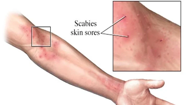
Scabies: The Intensely Pruritic Infestation
Scabies, caused by the mite Sarcoptes scabiei, creates one of the most intensely itchy conditions in dermatology and demonstrates high contagiousness through close personal contact.
Clinical Presentation: The hallmark symptom is intense nocturnal pruritus, occurring because mites are most active at night. Diagnostic burrows, though pathognomonic, may be difficult to identify due to scratching and secondary changes. The condition typically affects web spaces between fingers, wrists, palms, soles, genitalia, and axillae.
Diagnostic Considerations: Family members or close contacts often develop symptoms simultaneously, providing an important diagnostic clue. The condition should be suspected when multiple household members experience new-onset pruritus.
Treatment Protocol:
First-Line Topical Therapy:
- Permethrin 5% cream: Apply from neck down, leave for 8-14 hours, then wash off. Repeat application after 1 week.
- Coverage: Must treat entire body surface below the neck, including web spaces, genitalia, and under fingernails
Alternative Treatments:
- Ivermectin: orally, repeated after 1 week. Particularly useful for institutional outbreaks or treatment failures
- Benzyl benzoate 25%: Applied nightly for 3 consecutive nights
- Sulfur 6-10% ointment: Safe option for pregnant women and young children
Environmental Management:
- Wash all clothing and bedding in hot water (at least 50°C) and dry on hot cycle
- Items that cannot be washed should be sealed in plastic bags for 1 week
- Vacuum furniture and carpets, though environmental contamination is less important than direct contact
Treatment Success Factors: Simultaneous treatment of all household members and close contacts is essential to prevent reinfection. Itching may persist for several weeks after successful treatment due to residual immunologic reactions.
Fungal Infections: The Persistent Colonizers
Fungal infections of the skin represent a significant portion of dermatological practice, particularly in warm, humid climates that favor fungal growth.
Tinea Corporis and Cruris: The Ring-Makers
These superficial fungal infections affect the body (corporis) and groin area (cruris), caused by dermatophyte fungi that have adapted to digest keratin.
Clinical Recognition: The characteristic presentation involves circular, erythematous plaques with raised, scaly borders and central clearing. This “ring-like” appearance gives rise to the common name “ringworm,” though no worms are involved.
Common Causative Organisms:
- Trichophyton rubrum: Most common cause, often chronic and recurrent
- Trichophyton mentagrophytes: More inflammatory, often animal-acquired
- Microsporum canis: Usually acquired from infected pets
- Epidermophyton floccosum: Particularly affects groin area
Treatment Strategies:
Topical Therapy (first-line for localized disease):
- Terbinafine 1% cream: Apply twice daily for 2-4 weeks, continuing 1 week beyond clinical clearing
- Imidazoles: Clotrimazole, miconazole, or ketoconazole 1-2% applied twice daily for 2-4 weeks
- Treatment duration: Continue for 1-2 weeks after clinical resolution to prevent recurrence
Systemic Therapy (for extensive or resistant disease):
- Terbinafine
- Itraconazole
- Griseofulvin
Treatment Optimization: Success depends on adequate duration of therapy, addressing predisposing factors (humidity, tight clothing), and treating potential sources of reinfection (pets, family members).
Pityriasis Versicolor: The Color-Changing Condition
This superficial fungal infection, caused by Malassezia species, creates distinctive pigmentary changes that give the condition its name “versicolor” (many colors).
Clinical Features: The condition presents as sharply demarcated, hypopigmented or hyperpigmented macules and patches with fine scaling, predominantly affecting the upper trunk. The scaling becomes more apparent when the skin is stretched or scraped gently.
Pathophysiology: Malassezia yeasts are normal skin commensals that become pathogenic under certain conditions (heat, humidity, immunosuppression, hormonal changes). They produce azelaic acid, which inhibits melanin synthesis, leading to the characteristic hypopigmentation.
Treatment Options:
Topical Therapy:
- Ketoconazole 2% shampoo: Apply to affected areas, leave for 5 minutes, then rinse. Use daily for 1 week, then weekly for maintenance
- Selenium sulfide 2.5%: Apply at bedtime, wash off in morning. Use nightly for 1 week
- Terbinafine 1% solution: Apply twice daily for 1-2 weeks
Systemic Therapy (for extensive disease):
- Ketoconazole
- Itraconazole
- Fluconazole
Patient Education: Pigmentary changes may take several months to normalize after successful treatment. The condition commonly recurs, particularly in hot, humid conditions, and may require maintenance therapy.
Systemic Causes of Skin Symptoms
Not all skin problems originate in the skin itself. Generalized pruritus without obvious skin disease should prompt investigation for underlying systemic conditions.
Approach to Generalized Pruritus
Clinical Assessment: A systematic approach includes detailed history-taking, physical examination, and appropriate laboratory investigations to identify potential systemic causes.
Hematological Causes:
- Iron deficiency anemia: Can cause pruritus before other symptoms appear
- Polycythemia vera: Classic association with aquagenic pruritus (itching after bathing)
- Lymphomas: Particularly Hodgkin’s disease, may present with intractable itching
- Paraproteinemias: Multiple myeloma and related conditions
Hepatic Causes:
- Primary biliary cirrhosis: Often presents with pruritus years before jaundice
- Cholestasis: Any cause of bile acid accumulation can trigger intense itching
- Chronic liver disease: Advanced hepatic dysfunction from any cause
Renal Causes:
- Chronic kidney disease: Uremic pruritus affects up to 90% of dialysis patients
- Mechanism: Likely related to accumulation of uremic toxins and secondary hyperparathyroidism
Endocrine Causes:
- Hyperthyroidism: Increased metabolic rate and heat production
- Hypothyroidism: Associated with dry skin and pruritus
- Diabetes mellitus: Through multiple mechanisms including neuropathy and increased infection risk
Malignancies: Various cancers can present with paraneoplastic pruritus, sometimes preceding other symptoms by months or years.
Treatment Approach
Primary Treatment: Address the underlying systemic condition when identified. This often provides the most effective relief of pruritus.
Symptomatic Management:
- Antihistamines: H1 antagonists like cetirizine 10mg daily or loratadine 10mg daily
- Emollients: Regular application to maintain skin barrier function
- Topical anti-itch agents: Calamine lotion, menthol, or camphor-containing preparations
- Advanced therapies: For refractory cases, consider gabapentin, pregabalin, or tricyclic antidepressants
Prevention and Maintenance
Daily Skin Care Fundamentals
Cleansing Principles: Use lukewarm water and gentle, fragrance-free cleansers. Avoid hot water and harsh soaps that strip natural skin oils and disrupt the skin barrier.
Moisturizing Strategy: Apply moisturizers to slightly damp skin to trap moisture. Choose products appropriate for your skin type and environmental conditions.
Sun Protection: Daily use of broad-spectrum sunscreen with SPF 30 or higher prevents both acute damage and long-term complications including skin cancer.
Recognition of Concerning Changes
Warning Signs Requiring Medical Evaluation:
- New or changing moles, particularly those with asymmetry, irregular borders, color variation, or diameter >6mm
- Persistent sores that don’t heal within 2-3 weeks
- Rapidly growing or changing lesions
- Unexplained skin symptoms lasting longer than expected for common conditions
When to Seek Professional Care
Immediate Medical Attention:
- Signs of serious bacterial infection (fever, red streaking, rapid spread)
- Severe allergic reactions
- Extensive or rapidly spreading rashes
- Skin symptoms in immunocompromised patients
Routine Dermatological Care:
- Annual skin examinations for those at high risk for skin cancer
- Evaluation of persistent or recurrent skin problems
- Professional assessment of suspicious lesions
Conclusion
Understanding common dermatological conditions empowers individuals to make informed decisions about their skin health. While this comprehensive guide provides valuable insights into recognition and treatment options, it serves as an educational resource rather than a substitute for professional medical care.
The key principles of successful skin health management include early recognition of problems, appropriate use of treatments, addressing underlying predisposing factors, and knowing when to seek professional help. Many skin conditions can be prevented through proper hygiene, sun protection, and general health maintenance.
Remember that skin health reflects overall well-being, and persistent or unusual skin symptoms may indicate underlying systemic conditions requiring comprehensive evaluation. Building a relationship with qualified healthcare providers ensures access to the most current and appropriate treatments for your individual needs.
By understanding your skin’s language and responding appropriately to its signals, you can maintain optimal skin health throughout your life while recognizing when professional intervention becomes necessary.