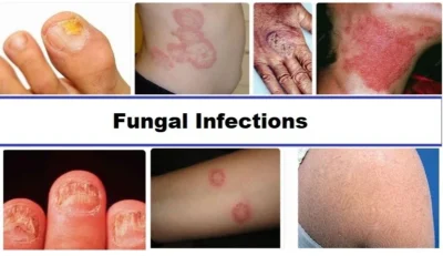
Comprehensive Guide to Superficial Fungal Infections
Introduction
Superficial fungal infections represent one of the most common dermatological conditions worldwide, affecting millions of people across all age groups and geographic regions. These infections, primarily caused by dermatophytes, yeasts, and other opportunistic fungi, affect the outermost layers of the skin, hair, and nails. According to the study, fungal skin diseases account for approximately 20-25% of all infectious skin conditions globally, with higher prevalence rates observed in tropical and subtropical regions where warm, humid climates create optimal conditions for fungal growth.
The clinical significance of superficial mycoses extends beyond mere cosmetic concerns, as these infections can cause substantial morbidity, psychological distress, and economic burden. A comprehensive understanding of their pathophysiology, clinical presentations, diagnostic approaches, and therapeutic management is essential for healthcare practitioners to provide effective patient care and prevent complications.
Dermatophytosis: The Ring of Infection
Etiology and Pathogenesis
Dermatophytosis, commonly known as ringworm, is caused by keratinophilic fungi belonging to three primary genera: Trichophyton, Microsporum, and Epidermophyton. These dermatophytes possess unique enzymatic capabilities that allow them to digest keratin, the primary structural protein found in the stratum corneum, hair, and nails. Research published in the Journal of Medical Microbiology indicates that dermatophytes secrete various keratinases and proteases that facilitate their invasion and survival within keratinized tissues.
The pathogenesis involves the initial adherence of fungal spores to the stratum corneum, followed by germination and hyphal invasion. The inflammatory response generated is primarily due to the permeation of fungal metabolic products and delayed-type hypersensitivity reactions. According to studies conducted by the International Society for Human and Animal Mycology, the host’s immune response plays a crucial role in determining the clinical presentation and severity of infection.
Clinical Classifications and Presentations
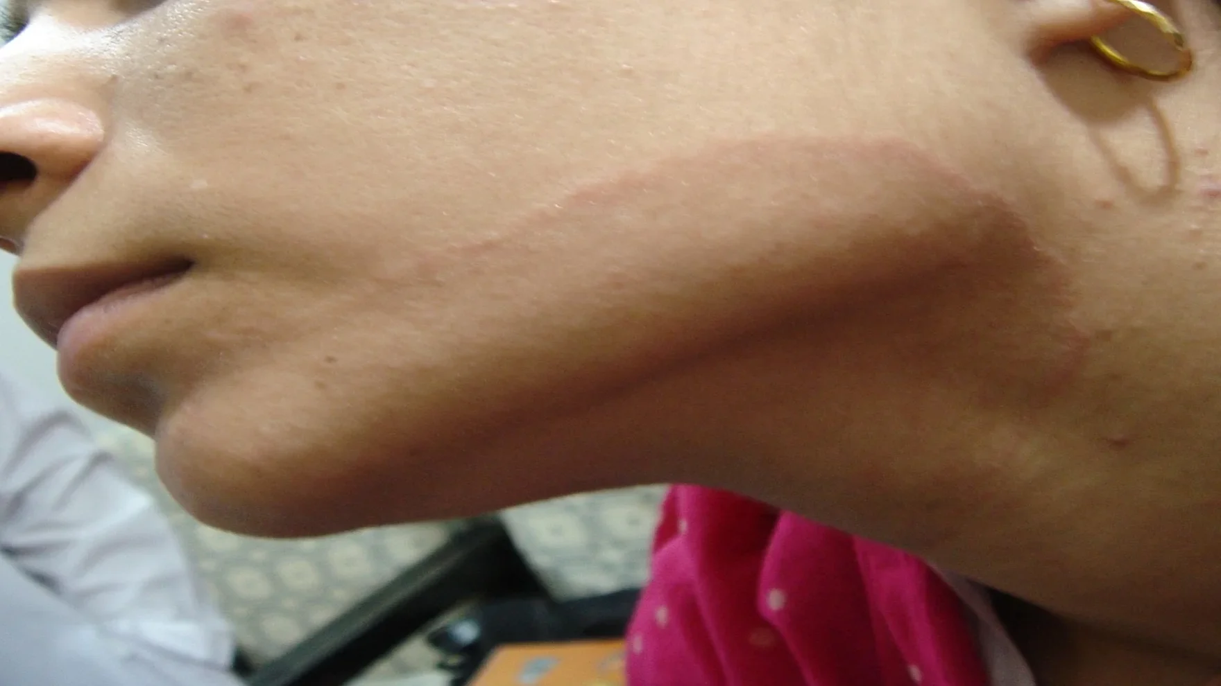
Tinea Corporis (Body Ringworm)
Tinea corporis manifests as the classic annular lesions with characteristic centrifugal spread. The typical lesion presents with a red, raised, scaly border and a relatively clear center, creating the distinctive “ring-like” appearance. The active edge shows papulo-vesicular changes, pustulation, and scaling, while the center may display hyperpigmentation, nodules, or thickening over time.
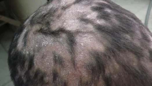
Tinea Capitis (Scalp Ringworm)
Predominantly affecting children, tinea capitis presents as patches of partial hair loss accompanied by scaling and sometimes black dots (broken hair shafts). The affected hair becomes lusterless and easily pluckable. According to pediatric dermatology studies, tinea capitis can manifest in both non-inflammatory and inflammatory forms, with the latter potentially leading to permanent scarring and alopecia if left untreated.
Tinea Cruris (Jock Itch)
This condition typically affects men more than women and is particularly common during summer and rainy seasons. The infection involves the groin, inner thighs, and sometimes the scrotum, presenting as bilateral erythematous, scaly plaques with well-demarcated borders. Clinical research published in the American Journal of Dermatology shows that tinea cruris has a high recurrence rate due to predisposing factors such as excessive sweating and occlusive clothing.
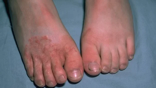
Tinea Pedis (Athlete’s Foot)
Tinea pedis primarily affects the interdigital spaces, particularly the lateral toe webs, presenting with scaling, maceration, and erosions. The condition is facilitated by occlusive footwear, excessive foot sweating, and communal bathing facilities. Studies indicate that tinea pedis often serves as a reservoir for other dermatophyte infections and may predispose patients to bacterial superinfections.
Tinea Unguium (Onychomycosis)
Nail involvement typically affects few nails asymmetrically and is often associated with concurrent fungal infections of feet, groin, or hands. The condition presents with nail plate separation from the nail bed, discoloration, and deformity. Research from the International Journal of Dermatology suggests that onychomycosis accounts for approximately 50% of all nail disorders and significantly impacts patients’ quality of life.
Tinea Incognito: The Masked Infection
A particularly challenging variant is tinea incognito, which occurs when typical dermatophyte infections are modified by inappropriate topical steroid use. This condition presents with atypical morphology, loss of well-demarcated borders, and unusual clinical features that can complicate diagnosis and treatment.
Pityriasis Versicolor: The Color-Changing Infection
Pityriasis versicolor is caused by Malassezia furfur (Pityrosporum ovale), a lipophilic yeast that exists as a normal skin commensal. The transition from commensal to pathogenic state occurs due to various predisposing factors including hot and humid conditions, genetic predisposition, immunosuppression, and hormonal changes.
The infection typically presents as hypopigmented, scaly lesions over the upper trunk and neck, though hyperpigmented and erythematous variants may also occur. According to dermatological surveys, pityriasis versicolor shows seasonal variation with higher incidence during warm, humid months. The condition is often asymptomatic but may cause mild itching and burning sensations.
Differential diagnosis primarily includes vitiligo, which presents with depigmented (not hypopigmented) lesions without scaling. Wood’s lamp examination may reveal characteristic fluorescence, and KOH preparation shows the pathognomonic “spaghetti and meatballs” appearance of short hyphae and round spores.
Candidiasis: The Opportunistic Invader
Pathophysiology and Predisposing Factors
Candida albicans, the primary causative organism of cutaneous candidiasis, exists as a normal commensal in the oral cavity, gastrointestinal tract, and genitourinary system. The transformation from commensal to pathogenic state occurs when the delicate host-microorganism balance is disrupted.
Key predisposing factors include:
- Moisture and occlusive conditions
- Diabetes mellitus
- Immunocompromised states
- Antibiotic therapy
- Pregnancy
- Obesity
Research published in Clinical Microbiology Reviews indicates that Candida species demonstrate remarkable adaptability, with the ability to switch between yeast and hyphal forms depending on environmental conditions.
Clinical Manifestations
Oral Thrush
Oral candidiasis presents as white, adherent plaques on an erythematous base affecting the buccal mucosa, tongue, and palate. The plaques can be wiped away, revealing raw, bleeding surfaces underneath. According to studies in immunocompromised patients, oral thrush often serves as an early indicator of systemic immune dysfunction.
Flexural Candidiasis
This variant affects skin folds including inframammary areas, axillae, groin, and finger webs. The characteristic presentation includes moist, red, macerated lesions in the depth of folds with satellite lesions extending beyond the main area of involvement. The satellite lesions are pathognomonic for candidal infection and help differentiate it from other intertrigenous dermatoses.
Candidal Paronychia
Nail fold involvement results in red, swollen periungual tissue with secondary nail plate changes including yellowing and deformity. Unlike bacterial paronychia, candidal paronychia typically develops gradually and may affect multiple fingers, particularly in individuals with frequent water exposure.
Genital Candidiasis
Vulvovaginal candidiasis presents with severe pruritus and characteristic white, curdy vaginal discharge. Male genital involvement manifests as superficial erosions and red, raw areas on the glans penis and prepuce.
Diagnostic Approaches
Laboratory Investigations
KOH (Potassium Hydroxide) Microscopy
KOH preparation remains the cornerstone of rapid fungal diagnosis. For dermatophytes, the examination reveals branching, septate hyphae and arthroconidia. Candida shows budding yeasts with pseudohyphae, while Malassezia demonstrates the characteristic “spaghetti and meatballs” pattern.
Fungal Culture
Culture remains the gold standard for species identification and antifungal sensitivity testing. Dermatophytes are typically cultured on Sabouraud’s dextrose agar at room temperature, with identification based on colony morphology and microscopic features.
Advanced Diagnostic Methods
Molecular techniques including PCR and DNA sequencing are increasingly being utilized for rapid and accurate species identification, particularly in cases where traditional methods yield inconclusive results.
Therapeutic Management
Topical Therapy
Antifungal Agents
- Imidazole derivatives: Miconazole, clotrimazole, ketoconazole, and econazole demonstrate broad-spectrum activity against dermatophytes, yeasts, and some non-dermatophyte molds.
- Allylamines: Terbinafine shows excellent activity against dermatophytes and is particularly effective due to its fungicidal mechanism of action.
- Polyenes: Nystatin remains effective against Candida species but lacks activity against dermatophytes.
Systemic Therapy
Terbinafine
This allylamine antifungal demonstrates fungicidal activity with excellent penetration into keratinized tissues.
Itraconazole
This triazole antifungal offers broad-spectrum activity against dermatophytes, yeasts, and some molds. Pulse therapy regimens have shown particular efficacy in onychomycosis treatment, with reduced side effects compared to continuous therapy.
Fluconazole
Particularly effective against Candida and Malassezia species, fluconazole offers excellent bioavailability and tissue penetration. Single-dose therapy (150mg) is often sufficient for uncomplicated vulvovaginal candidiasis.
Griseofulvin
Although still used in some settings, griseofulvin has largely been superseded by newer antifungals due to its fungistatic nature, long treatment duration requirements, and potential for drug interactions.
Prevention and General Management
Effective management of superficial fungal infections requires addressing predisposing factors and implementing preventive measures:
- Environmental modifications: Maintaining dry conditions, ensuring adequate ventilation, and avoiding occlusive garments
- Personal hygiene: Regular cleansing, thorough drying of skin folds, and avoiding sharing of personal items
- Medical management: Controlling underlying conditions such as diabetes mellitus and addressing immunosuppression
- Contact management: Treating family members and close contacts in cases of highly contagious infections like tinea capitis
Treatment Challenges and Resistance
According to recent studies published in Antimicrobial Agents and Chemotherapy, antifungal resistance is emerging as a significant concern, particularly with Candida species. Risk factors for treatment failure include:
- Inadequate treatment duration
- Poor medication compliance
- Failure to address predisposing factors
- Immunocompromised status
- Biofilm formation in chronic infections
Conclusion
Superficial fungal infections continue to represent a significant global health burden, affecting individuals across all demographics. The diversity of causative organisms, varied clinical presentations, and potential for complications underscore the importance of accurate diagnosis and appropriate treatment selection.
Healthcare providers must maintain a high index of suspicion for these infections, particularly in predisposed populations. Early recognition, proper diagnostic evaluation, and targeted therapy can significantly reduce morbidity and prevent transmission to close contacts.
The emergence of antifungal resistance highlights the need for antimicrobial stewardship and continued research into novel therapeutic approaches. Additionally, patient education regarding prevention strategies and treatment compliance remains crucial for successful outcomes.
As our understanding of fungal pathogenesis continues to evolve, personalized treatment approaches based on individual risk factors, causative organisms, and host immune status may further improve therapeutic outcomes and reduce the global burden of superficial mycoses.